Images Of Fetal Brain Activity Never Seen Before

Some time ago it was very difficult to get pictures of the brain activity of a fetus in the womb. Nowadays, thanks to highly developed technology, there are already high quality images. These enable us to better understand certain aspects of the development of a new human life that were previously unknown to us.
Magnetic resonance imaging (MRI) of the brain of a fetus is a diagnostic procedure that supplements ultrasound. V M any expectant mothers opt for a very specific reason:
“Life is fascinating, we just have to look at it from the right angle.”
Alejandro Dumas
These tests are usually done from the 20th week of pregnancy. At this point the corpus callosum has completely formed and the diagnosis is more accurate. We must remember that the fetus is surrounded by amniotic fluid. His world consists mainly of liquid, which means that magnetic resonance imaging does not provide high-resolution images. Because every movement makes a clear picture impossible.
This type of perinatal abnormality test usually has a 50% success rate. But now there are completely new recording methods. Not that long ago, technology took a giant leap forward, and much more accurate algorithms already exist that can take near-perfect readings of a baby’s brain activity.
What was discovered in these early diagnostic tests was a revolutionary discovery in the field of perinatal medicine. We’ll tell you about it below.
The brain activity of fetuses
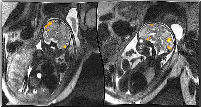
In the picture above we can see magnetic resonance imaging of a fetus at 20 weeks and another at 40 weeks of pregnancy. These images were taken from the Medical School of Wayne State University (Michigan, USA). On them we can clearly see the brain activity of these two babies in the womb.
With this research method, the scientists wanted to study how the neurons of the fetuses connect during the last weeks of pregnancy. The data obtained have revealed certain aspects relating to premature babies that were unknown up to that point.
Less cerebral connections in premature babies
The results of this first study were published in the journal ” Scientific Reports “. In order to carry out the said examination using the new magnetic resonance technology, half of these women had a high-risk pregnancy and subsequently a premature birth.
- The premature babies were found to have much weaker neural connections than other fetuses in the same week of pregnancy.
- Up until that point, it was thought that the lower cerebral connections found in premature babies were primarily due to dramatic childbirth or hypoxia that they often experience during childbirth.
However, this new diagnostic procedure revealed that the small cerebral connections are already evident in the womb and that this smaller connection between the neurons can mainly be seen in Broca’s area. This is the area of the brain that is related to language processing.
How useful will these new research methods be?
As we mentioned at the beginning, fetal magnetic resonance aims to detect any perinatal abnormality. Nowadays it is very noticeable that there are more and more premature babies. This fact obliges doctors, scientists and families to provide new strategies, forces and resources.
- The data obtained from this study have shown us that many of these premature babies in the womb are surrounded by inflamed placental tissue. Hence, scientists believe
- The earlier these perinatal anomalies are discovered, the easier it is to intervene in good time. We must keep in mind that premature babies are at greater risk of developing autism, attention deficits, and other special learning needs.
“When you least expect it, life presents you with a challenge.”
Paulo Coelho
In summary, we can say that these first pictures of the brain activity of human fetuses show us extraordinary things, which allow us to understand the development of humans a little better. But above all, this examination method will in the future be a very precise diagnostic procedure with which a premature baby, which science, doctors and his family needs so much , can be examined more completely.
At least we hope it will.