Research Methods In The Field Of Biopsychology
Authors such as Dewsbury (1991) define biopsychology as “the scientific study of the biology of behavior”. This field is also known as psychobiology. However, other authors prefer the term “biopsychology” because it represents “a biological approach to the study of psychology, rather than a psychological approach to the study of biology”.
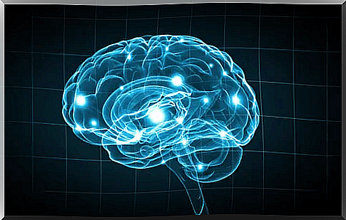
Thanks to scientific advances, research methods in the field of biopsychology have experienced an enormous revolution in recent years: Countless older researchers thought they would be able to observe brain functions in real time at some point. And today it is possible. In general, the focus is on those methods that allow knowledge about what happens in the brain under certain conditions.
“Man is the most mysterious and disturbing scientific discovery.”
Ganivet
Imaging diagnostics
Observing and recording brain activity in real time is a milestone achieved thanks to the various techniques developed over the course of the twentieth century. These are techniques that undoubtedly represent a great step forward when it comes to the organ that remains the least explored.
X-rays and contrast media
In this technique, a substance that absorbs X-rays is injected into a part of the body, into a specific system. This increases the contrast between the body part to be examined and the surrounding tissue.
For example, cerebral angiography is an X-ray procedure that uses contrast media. To do this, an opaque dye is injected into a cerebral artery. The aim is to observe the cerebral circulation during the X-ray. This technique is useful in localizing vascular lesions and brain tumors.
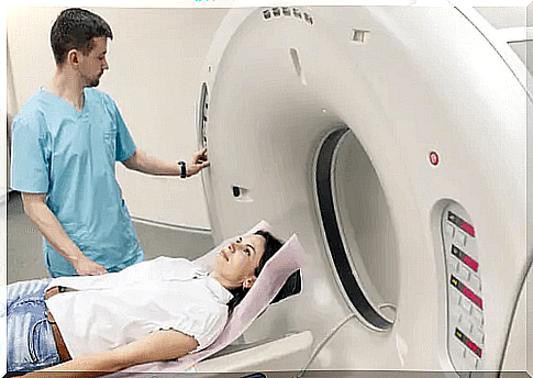
Computed tomography (CT)
The brain structure can be illustrated in detail in the CT. During the examination, the patient lies in a tube provided for this purpose. When lying down, the X-ray tube and a receiver, which are diametrically opposite each other, take many individual images. This happens while the transmitter and receiver circle around the patient’s head.
The information from all the images created is merged with the help of a computer. This unification makes it possible to explore the brain in levels. Usually this exam is done in eight or nine layers of the brain. Once all the images have been combined, you get a three-dimensional representation of the brain.
Nuclear Magnetic Resonance or Magnetic Resonance (NMR / MRT)
The NRM / MRI provides high resolution images that show the movement of hydrogen molecules when activated by high frequency waves in a magnetic field. It enables a high spatial resolution and also generates three-dimensional images.
Positron Emission Tomography (PET)
PET scans provide images of brain activity, but not of the brain structure itself. To obtain these images , radioactive 2-deoxyglucose (2-DG) is injected into the patient’s carotid artery. The active neurons quickly absorb the 2-DG, and since it is not metabolized by them, the 2-DG accumulates until it is gradually broken down afterwards. In this way it can be observed which neurons are active at a certain point in time.
Functional magnetic resonance imaging (fMRI)
The fMRI creates images of the oxygen supply via the blood that flows through the individual brain regions. This also allows brain activity to be measured. This examination method has four advantages over PET:
- Nothing is injected into the patient.
- It provides both functional and structural information.
- It offers better spatial resolution.
- Three-dimensional images of the entire brain can be created.
Magnetoencephalography
It measures changes in magnetic fields that appear on the surface of the scalp. These changes, in turn, are caused by changes in the patterns of neural activity.
Transcranial Magnetic Stimulation (TMS)
Walsh and Rothwell (2000) explain that TMS is a technique that changes the activity of an area of the cortex and creates a magnetic field under a hood that sits on the head. The TMS temporarily deactivates part of the brain while the effects of this deactivation on behavior and cognition are assessed.
Harmful methods
Harmful research methods include those that destroy an area of the brain to find out how it affects behavior.
- Aspiration injuries. This method is usually used to create a lesion in certain areas of the brain that are visible to the naked eye. The corresponding tissue is removed with a very fine crystal pipette.
- High frequency lesions. These are small subcortical lesions. A high-frequency current is passed through the tissue to be destroyed with an electrode. The size and shape of the lesion depends on three factors:
- duration
- Intensity of the current
- Configuration of the electrode tip
- Scalpel cuts. It is cut into the area of the brain that is to be destroyed.
- Cold blockers. This procedure is reversible. Instead of destroying a structure, an area is cooled down until it reaches freezing point. Neurons thus stop sending signals and block the function of the cooled brain region. In this way it can be observed which behaviors are induced by the areas in which the intervention was made. Once the temperature returns to normal, normal brain function can also be restored.
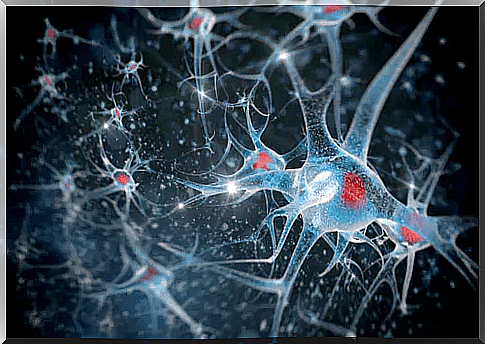
Electrical stimulation
Another research method in biopsychology is electrical stimulation. A structure of the nervous system is electrically stimulated in order to obtain information about its function. Usually a bipolar electrode is used for this.
This stimulation affects the neurons and changes their state. This usually has the opposite effect of lesions. For example, if an injury causes someone to drastically reduce their sleep times, stimulation can lead to a disproportionate need for sleep.
Harmful examination methods with electrophysiological recording
- Intracellular record of a unit. This technique is performed by inserting a microelectrode into a neuron. It provides a record of the graduated fluctuations in the neuron’s membrane potential.
- Extracellular record of a unit. Here the microelectrode is inserted into the extracellular fluid surrounding the neuron.
- Multiple unit recording. In this case, the tip of the electrode is larger than that of a microelectrode, so it can record signals from many neurons at the same time. The identified potentials are added and integrated.
- Invasive EEG recording. In this case the electrodes are implanted. When searching for cortical EEG signaling protocols, cranial “mother electrodes” made of stainless steel are used. Cable electrodes that are implanted in a stereotactic procedure are often used for subcortical signals.
“Anthropology, biology, physiology and psychology have gathered mountains of materials in order to establish the foundations of their physical and mental perfection and their further development in the face of all human facets.”
Leon Trotsky
Research methods in the field of biopsychology: There is still a long way to go
In the course of the article we have explained some of the bio-psychological research methods. However, we do not want to leave it unsaid that there are other methods in biopsychology that examine other areas of the body. This includes measuring muscle tension, recording eye movements, skin conductivity or cardiovascular activity.
The progress that has recently been made in the investigative methods of biopsychology is remarkable, but it has not come to an end. In other words, in a few years time, new techniques are sure to emerge that we cannot even imagine right now. All of this will help advance neuroscience, which in turn will help improve the quality of life for many people affected by a nervous system disorder.